

We deliver specialized, patient centered orthopedic care combining advanced surgical techniques with cutting edge regenerative medicine. Our practice is dedicated to accurately diagnosing joint conditions, restoring natural biomechanics, and providing personalized treatment plans that help you return to an active, pain free life

SERVICES
1. SHOULDER SURGERY
The shoulder is the most mobile joint in the human body, relying on a complex network of muscles, tendons, and ligaments for stability. When non-surgical treatments fail to resolve chronic pain, instability, or structural tears, specialized surgical procedures can restore full function and overhead mobility.
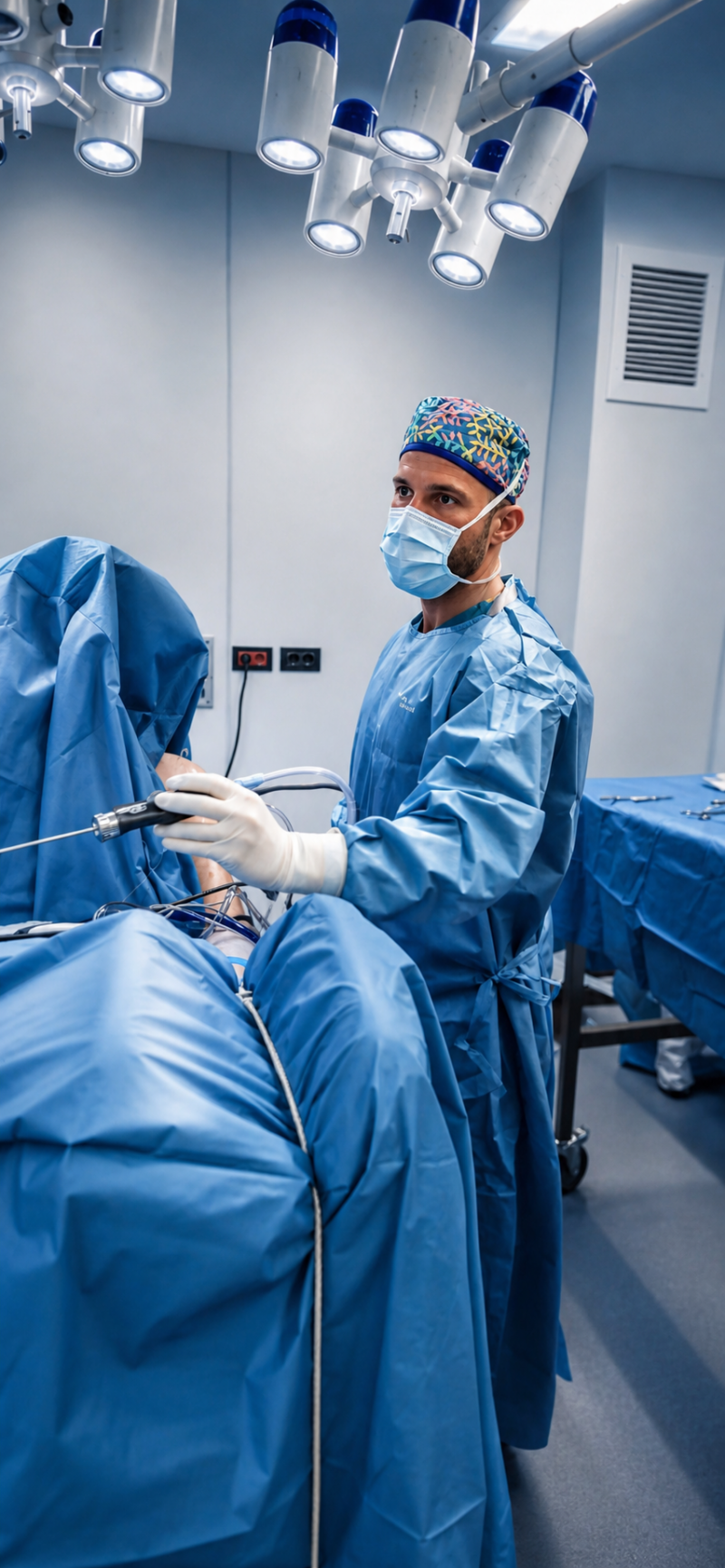
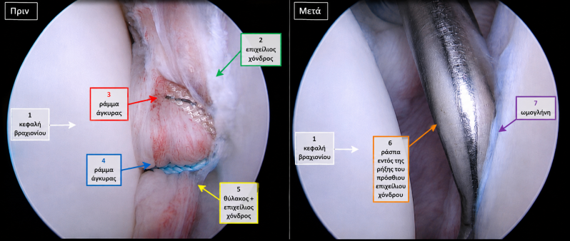
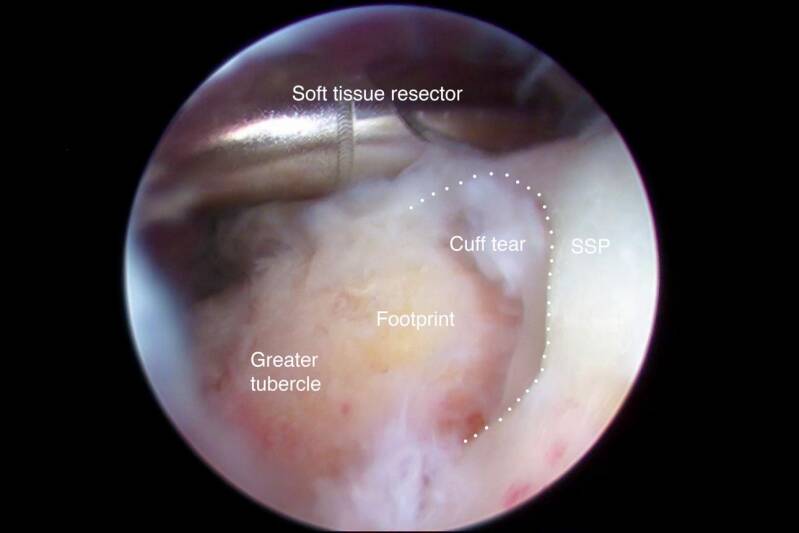
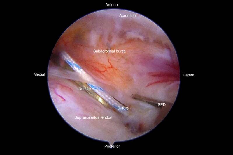
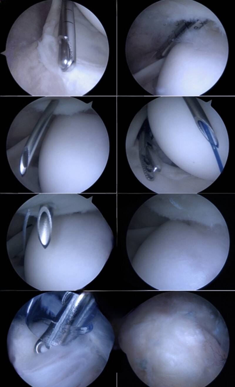
A. Arthroscopic Rotator Cuff Repair
What It Is:
A minimally invasive procedure aimed at reattaching torn tendons of the rotator cuff—specifically the supraspinatus, infraspinatus, subscapularis, or teres minor—to the head of the humerus (the upper arm bone).
How the Procedure Works:
Incisions & Portal Creation: Small, pencil-sized incisions (portals) are made around the shoulder joint.
Arthroscopic Visualization: A high-definition camera (arthroscope) is inserted, enabling the surgeon to inspect the rotator cuff, joint lining, and surrounding cartilage in real time.
Bone Bed Preparation & Anchor Placement: The damaged bone area is cleaned to facilitate healing, and specialized suture anchors, made from biocompatible material or titanium, are inserted into the humerus.
Tendon Reattachment: Sutures from the anchors are woven through the torn tendon and tightened, securely reattaching the rotator cuff to the bone.
Common Indications:
Full-thickness or significant partial-thickness rotator cuff tears, persistent shoulder weakness, night pain, and difficulty raising the arm.
Recovery Timeline:
A sling is worn for 4 to 6 weeks to protect the tendon repair, followed by a structured physical therapy program. Full athletic recovery and maximum strength typically take 6 to 9 months.
B. Labral Repair & Instability Surgery (Bankart & SLAP Repair)
What It Is:
Surgeries designed to repair tears in the glenoid labrum—the cartilage ring that deepens the shoulder socket and anchors key ligaments and the biceps tendon.
How the Procedure Works:
Bankart Repair (Anterior Instability): This procedure is performed when the labrum detaches from the front of the socket, often due to shoulder dislocation. Sutures and small anchors are used to reattach the labrum and tighten the stretched joint capsule.
SLAP Repair (Superior Labrum Anterior to Posterior): This addresses tears at the top of the socket where the long head of the biceps tendon attaches. The torn labrum is cleaned and reattached using suture anchors.
Latarjet Procedure (Open Reconstruction): For cases of severe bone loss or recurrent dislocations, a small piece of bone with its attached tendon (the coracoid process) is transferred to the front of the shoulder socket to rebuild missing bone and prevent further dislocations.
Common Indications:
Recurrent shoulder dislocations, sensations of "catching" or "clicking," deep joint pain in overhead throwers, and traumatic sports injuries.
Recovery Timeline:
A sling is used for 4 weeks, with range-of-motion exercises beginning early. Overhead sports and heavy lifting can typically be resumed 5 to 7 months post-surgery.
C. Total & Reverse Shoulder Replacement (Arthroplasty)
What It Is:
A surgical procedure that replaces worn-out or severely damaged shoulder joint surfaces with durable prosthetic components made of metal and medical-grade polyethylene plastic.
How the Procedure Works:
Anatomical Total Shoulder Replacement: This approach restores the natural anatomy. The arthritic ball (humeral head) is replaced with a smooth metal sphere, and the worn socket (glenoid) is lined with a smooth plastic cup. This procedure requires a healthy, functioning rotator cuff.
Reverse Total Shoulder Replacement: This technique reverses the positions of the ball and socket. The metal ball is attached to the shoulder blade (socket side), while the plastic socket is affixed to the arm bone (ball side). This allows the deltoid muscle to lift the arm instead of the torn rotator cuff.
Common Indications:
Severe osteoarthritis, rheumatoid arthritis, complex multi-fragment shoulder fractures, and rotator cuff tear arthropathy (end-stage joint wear with irreparable tendon tears).
Recovery Timeline:
A hospital stay of 1 to 2 days is typical, with sling use for 3 to 4 weeks. Most patients experience significant pain relief within weeks, with functional range of motion restored over a period of 3 to 6 months.
2. KNEE SURGERY
The knee is a complex, weight-bearing hinge joint reliant on ligaments, cartilage, and menisci for stability and fluid movement. When injury or progressive degeneration limits mobility and causes chronic pain, targeted surgical interventions can repair damaged structures, correct alignment, and restore joint function.
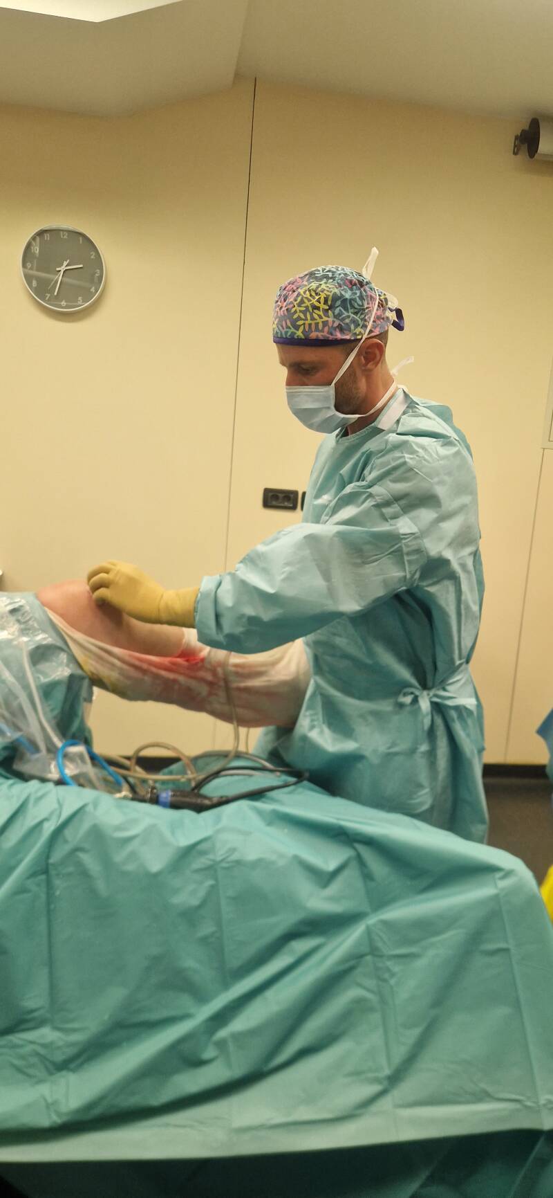
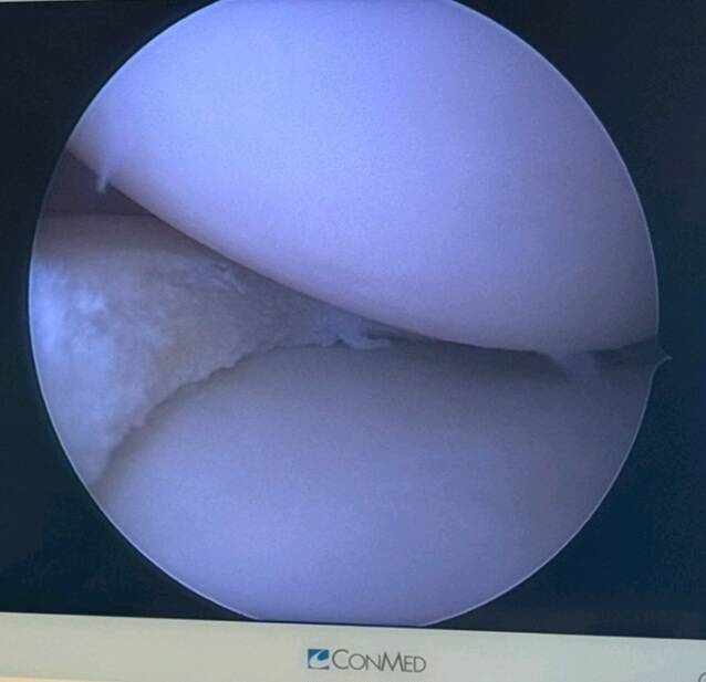
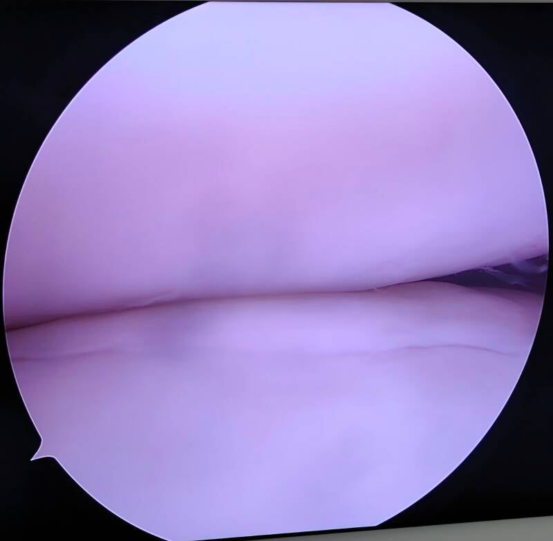
A. Anterior Cruciate Ligament (ACL) & Multi-Ligament Reconstruction
What It Is:
This surgical procedure involves the reconstruction of a torn Anterior Cruciate Ligament (ACL), a key stabilizer of the knee. The goal is to restore rotational stability and prevent the knee from buckling during high-impact activities.
How the Procedure Works:
Graft Preparation: Since a torn ligament cannot be stitched back together, a graft is selected and prepared. This graft can be an autograft (harvested from your own hamstring, patellar tendon, or quadriceps tendon) or an allograft (donor tissue).
Tunnel Drilling: Using minimally invasive arthroscopic techniques, precise tunnels are drilled into the tibia (shinbone) and femur (thighbone) at their anatomical attachment points.
Graft Fixation: The new ligament graft is threaded through the bone tunnels and securely anchored using specialized biocompatible screws, buttons, or posts.
Common Indications:
This procedure is indicated for complete ACL tears, complex multi-ligament knee injuries (such as PCL, MCL, LCL), recurrent knee instability, and for athletes engaged in high-demand activities requiring pivoting or cutting movements.
Recovery Timeline:
During the first 2 to 4 weeks, patients use crutches and a protective knee brace. Early physical therapy focuses on regaining full extension and activating the quadriceps. A full return to competitive sports typically requires 8 to 12 months of structured rehabilitation.
B. Meniscus Repair & Cartilage Restoration
What It Is:
Arthroscopic procedures aimed at preserving as much natural meniscal and articular cartilage tissue as possible. This approach helps delay joint degeneration and maintains smooth joint mechanics.
How the Procedure Works:
Meniscus Repair: For tears located in the outer "red zone" (which has a rich blood supply), specialized suture devices are utilized through small arthroscopic portals to stitch the torn edges together.
Partial Meniscectomy: If the tear is in an unvascularized region (the "white zone") or is severely frayed, the surgeon carefully trims away only the damaged portion, preserving as much healthy meniscus as possible.
Cartilage Restoration (Microfracture / OATS / ACI): Small holes are created in the bone (microfracture), or cartilage plugs are transferred (OATS/osteochondral grafting) to stimulate new tissue growth over cartilage defects.
Common Indications:
This procedure is appropriate for meniscal tears that cause joint locking or clicking, focal cartilage defects, localized knee pain, and persistent swelling resulting from sports injuries or acute trauma.
Recovery Timeline:
Partial Meniscectomy: Patients experience rapid recovery, with weight-bearing as tolerated within days and full recovery in 3 to 6 weeks.
Meniscus Repair / Cartilage Restoration: There are weight-bearing restrictions for 4 to 6 weeks to protect healing tissue; full athletic recovery typically occurs within 4 to 6 months.
C. Partial & Total Knee Arthroplasty (Knee Replacement)
What It Is:
This resurfacing procedure involves removing damaged cartilage and bone surfaces and replacing them with high-durability metallic components and medical-grade polyethylene inserts.
How the Procedure Works:
Partial (Unicompartmental) Knee Replacement: This option is used when arthritis is confined to a single compartment of the knee (either medial or lateral). Only the damaged compartment is resurfaced, preserving healthy cartilage, bone, and natural ligaments (including the ACL and PCL).
Total Knee Replacement: This is indicated for widespread end-stage osteoarthritis. All three compartments of the joint are resurfaced using precision alignment instruments to ensure correct leg axis and implant longevity.
Common Indications:
Severe osteoarthritis, rheumatoid arthritis, post-traumatic arthritis, significant stiffness, bone-on-bone joint wear, and persistent pain that limits daily activities.
Recovery Timeline:
Patients typically begin walking on the day of surgery or the following morning. Most transition off assistive devices within 2 to 4 weeks. Major pain relief and a return to normal daily activities are generally achieved within 6 to 12 weeks, with continued structural improvement over the course of a year.
3. HIP SURGERY
The hip is a robust ball-and-socket joint designed to bear body weight and allow a wide range of fluid movement. Pain in the hip can stem from structural impingement, cartilage tears, or progressive wear-and-tear (osteoarthritis). Our specialized hip procedures focus on joint preservation for active individuals and advanced, muscle-sparing replacement techniques for end-stage joint damage.
A. Hip Arthroscopy & Joint Preservation
What It Is:
A cutting-edge, minimally invasive procedure that utilizes small incisions and specialized cameras to diagnose and treat intra-articular hip issues without the need for open surgery.
How the Procedure Works:
Joint Distraction: Gentle traction is applied to the leg, creating space in the hip socket for safe access with arthroscopic instruments.
Correction of Impingement (FAI):
CAM Correction: Bony overgrowths at the junction of the femoral head and neck are reshaped (femoroplasty) to achieve a smooth, spherical contour.
Pincer Correction: Excess bone extending over the rim of the acetabulum (socket) is trimmed back (acetabuloplasty).
Labral Repair & Cartilage Debridement: The torn labrum, which serves as a rubbery seal around the socket, is anchored back to the bone using bio-absorbable sutures. Damaged articular cartilage is smoothed or repaired.
Common Indications:
Femoroacetabular Impingement (FAI), acetabular labral tears, loose bodies within the joint, painful hip clicking or "catching," and early cartilage lesions in young or active individuals.
Recovery Timeline:
Patients typically use crutches for 2 to 4 weeks to protect the repaired tissue. A structured physical therapy program focuses on restoring motion and strengthening the core and glutes. Most patients can return to sports and resume unrestricted activities between 4 and 6 months.
B. Muscle-Sparing Total Hip Replacement (Direct Anterior & Modern Posterior)
What It Is:
This procedure involves surgically replacing damaged components of the hip joint with highly durable, biocompatible implants (titanium alloy, highly cross-linked polyethylene, or ceramic) to alleviate severe pain and restore mobility.
How the Procedure Works:
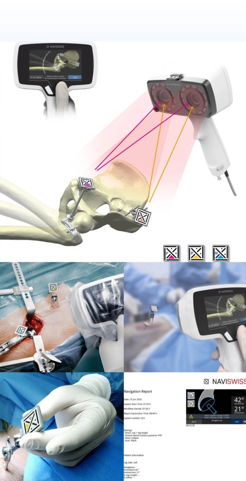
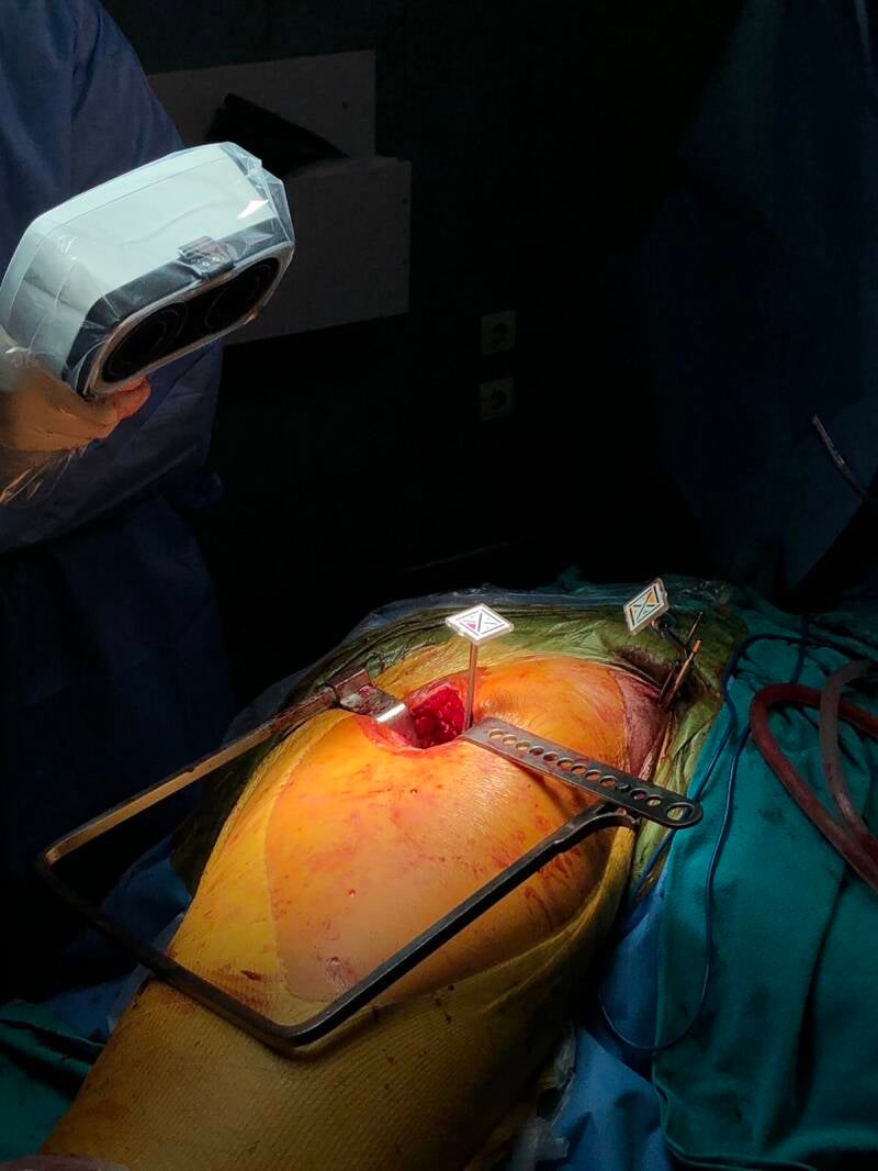
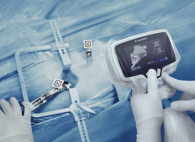
Joint Exposure: Utilizing modern, minimally invasive techniques, such as the Direct Anterior Approach (entering from the front without detaching or cutting muscles) or specialized soft-tissue-preserving posterior techniques.
Removal of Diseased Bone: The worn femoral head is removed, and the arthritic socket is gently reamed to clean the bone.
Prosthetic Implant Placement: A durable metal socket with a ceramic or plastic liner is securely fixed into the pelvis, and a metal stem with a polished ceramic ball is inserted into the femur.
Common Indications:
Advanced osteoarthritis, avascular necrosis (loss of blood supply to the hip bone), rheumatoid arthritis, severe post-traumatic joint wear, and debilitating pain that limits walking, sitting, or sleeping.
Recovery Timeline:
Most patients are encouraged to stand and walk with a walker or crutches on the day of surgery or the following morning. Most transition off walking aids within 1 to 3 weeks and can resume light daily activities, driving, and swimming within 3 to 6 weeks.
C. Revision Hip Surgery (Complex Reconstructive Arthroplasty)
What It Is:
A specialized and complex surgical procedure aimed at replacing a previously implanted artificial hip joint that has failed over time due to wear, loosening, dislocation, or infection.
How the Procedure Works:
Explantation: The failed or loose implants are carefully removed while preserving as much surrounding bone stock as possible.
Bone Reconstruction: If bone loss has occurred around the joint, specialized bone grafts or porous titanium metal augments are utilized to rebuild the socket and femur.
Revision Implant Placement: Custom, modular, or specialized long-stem implants are inserted to ensure stable, long-lasting fixation.
Common Indications:
Mechanical loosening of old implants, severe implant wear, recurrent dislocations, deep joint infection around a prosthesis, or periprosthetic fractures (fractures around the existing implant).
Recovery Timeline:
Recovery duration varies based on the complexity of the reconstruction. Patients often use crutches or a walker for 6 to 12 weeks, adhering to customized weight-bearing restrictions while new bone integrates with the specialized implants. Full recovery typically spans 6 to 12 months.
4. BIOLOGICAL THERAPIES
Biological therapies, also known as regenerative medicine, harness your body’s natural healing processes to alleviate pain, diminish inflammation, and speed up tissue repair. These non-surgical, minimally invasive treatments are conducted in an outpatient setting, providing an effective alternative or complement to traditional orthopedic procedures.
A. Platelet-Rich Plasma (PRP) Therapy
What It Is:
PRP therapy harnesses concentrated platelets derived from your own blood. These platelets are rich in active growth factors—proteins vital for cellular signaling, blood vessel formation, and tissue regeneration.
How the Procedure Works:
Blood Collection: A standard blood sample (approximately 10–20 mL) is drawn from your arm.
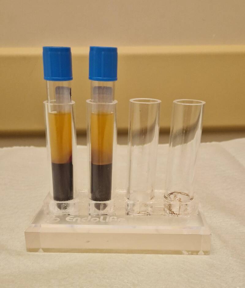
Centrifugation: The blood is placed in a high-speed centrifuge for 10–15 minutes, effectively isolating the growth-factor-rich plasma from red and white blood cells.
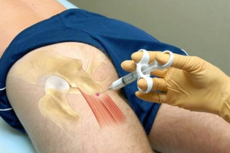
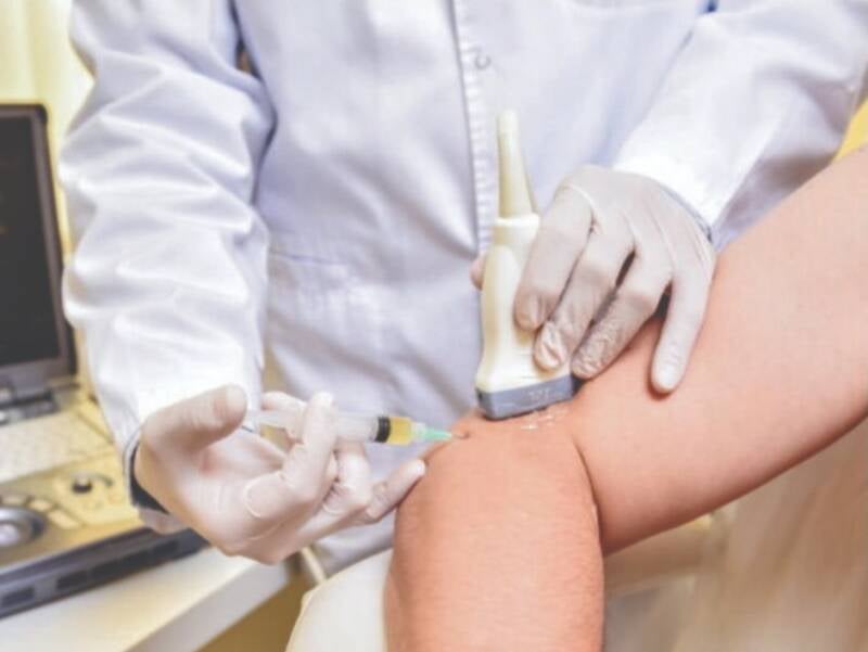
Targeted Ultrasound-Guided Injection: The concentrated PRP is precisely injected into the affected tendon, ligament, or joint space using real-time ultrasound guidance to ensure accurate delivery.
Common Applications:
PRP therapy is commonly used for conditions such as rotator cuff tendinopathy, tennis and golfer’s elbow, patellar tendinitis, Achilles tendinitis, plantar fasciitis, and mild-to-moderate joint osteoarthritis.
Recovery & Results:
Patients often experience localized soreness for 2–5 days as the acute inflammatory healing process begins. Significant pain relief and functional improvement generally manifest over a period of 4 to 8 weeks.
B. Cellular & Bone Marrow Aspirate Concentrate (BMAC) Therapies
What It Is:
BMAC therapy provides a rich blend of adult mesenchymal stem cells, progenitor cells, and anti-inflammatory cytokines directly to damaged cartilage and joint structures.
How the Procedure Works:
Harvesting: Under local anesthesia and light sedation, a specialized needle is used to aspirate a small amount of bone marrow, typically from the posterior hip or iliac crest.
Processing: The aspirated bone marrow is processed in a specialized centrifuge system to concentrate the regenerative cellular elements.
Precision Delivery: The final biological concentrate is injected directly into the affected joint or osteochondral lesion under live guidance (ultrasound or fluoroscopy).
Common Applications:
BMAC therapy is effective for early-to-moderate osteoarthritis of the knee, hip, or shoulder, focal cartilage defects, non-healing bone fractures, and persistent soft tissue tears.
Recovery & Results:
Patients experience minimal downtime, with restricted high-impact activities for 1–2 weeks. Cell-mediated repair and matrix reorganization occur gradually, providing long-term structural support and pain relief over 3 to 6 months.
C. Viscosupplementation (Hyaluronic Acid Injections)
What It Is:
Hyaluronic acid (HA) is a naturally occurring component of healthy synovial fluid, functioning as a lubricant and shock absorber within joint cavities. In osteoarthritic joints, the body’s natural HA degrades, leading to increased joint friction, stiffness, and pain.
How the Procedure Works:
Preparation: The skin over the joint is thoroughly cleansed, and a local anesthetic is applied to minimize discomfort.
Joint Aspiration (If needed): Any excess fluid accumulation (joint effusion) is drained first to relieve pressure.
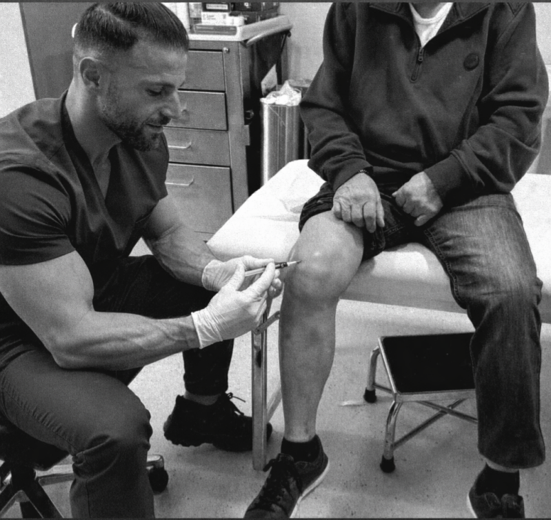
Injection: A high-molecular-weight hyaluronic acid formulation is injected directly into the joint capsule, quickly restoring fluid viscosity and mechanical shock absorption.
Common Applications:
This treatment is typically recommended for mild-to-moderate osteoarthritis of the knee, hip, or shoulder, especially for patients who cannot tolerate non-steroidal anti-inflammatory drugs (NSAIDs) or wish to postpone joint replacement surgery.
Recovery & Results:
Patients can resume light daily activities immediately. Pain relief and reduced joint stiffness usually last between 6 and 12 months, and the treatment can be safely repeated as needed.